Hip And Leg Bone Diagram - Femur And Pelvic Bones Diagram Fusebox And Wiring Diagram Series Ton Series Ton Sirtarghe It - The ball and socket bony structure.
Hip And Leg Bone Diagram - Femur And Pelvic Bones Diagram Fusebox And Wiring Diagram Series Ton Series Ton Sirtarghe It - The ball and socket bony structure.. The ball and socket bony structure. Cited after worker's leg amputated. bones of the lower limb anatomy and physiology i these pictures of this page are about:leg bones diagram. The hip bone (os coxae, innominate bone, pelvic bone or coxal bone) is a large irregular bone, constricted in the center and expanded above and below. The medial muscles of the hip are involved in the adduction of the leg i.e. The femur is the upper leg bone or thigh.
Cited after worker's leg amputated. bones of the lower limb anatomy and physiology i these pictures of this page are about:leg bones diagram. Click and start learning now! Quizzes on human skeletal system anatomy, bone the musculoskeletal segment of the leg, including the foot bones (ankle, heel bone, toe bones), fibula and tibia, knee, femur and femoral neck, hip and sacrum. 12 photos of the diagram of leg bones. The pelvic bone area is different in men and women.
Hip Joint Anatomy Hip Bones Ligaments Muscles from bonesmart.org Learn about the hip joint, with its remarkable combination of strength and flexibility, using our interactive anatomy image it bears our body's weight and the force of the strong muscles of the hip and leg. By natalia kremenon january 21, 2021in wiring diagram231 views. The head of your femur fits into your hip socket and the bottom end connects to your knee. It is usually often called the calf bone, because it sits barely behind the tibia on the surface of the leg. Hip and thigh bones joints muscles kenhub. Bones of the hip joint. Quizzes on human skeletal system anatomy, bone the musculoskeletal segment of the leg, including the foot bones (ankle, heel bone, toe bones), fibula and tibia, knee, femur and femoral neck, hip and sacrum. Want to learn more about it?
Cited after worker's leg amputated. bones of the lower limb anatomy and physiology i these pictures of this page are about:leg bones diagram.
The medial muscles of the hip are involved in the adduction of the leg i.e. This bone attaches to the sacrum (forming the sacroiliac joint) and to its counterpart at the pubic symphysis, forming the pelvic girdle. Bones of the hip joint. Hip anatomy pictures function problems treatment 28 labeled diagram of the femur long bone diagram labeled The hip bone (os coxae, innominate bone, pelvic bone or coxal bone) is a large irregular bone, constricted in the center and expanded above and below. Bones of the hip joint. By natalia kremenon january 21, 2021in wiring diagram231 views. Use the leg bones diagrams to learn the names of the leg bones and leg anatomy. This lengthy bone connects with the knee at one finish and the ankle on the different. The head of your femur fits into your hip socket and the bottom end connects to your knee. Bones of the hip diagram identification 17 6 petraoberheit de lamb leg bones diagram 19 6 asyaunited de best anatomy of the thigh hip and pelvis femur diagram femoral vein muscles of the thigh anterior medial posterior teachmeanatomy. The hip joint is the uppermost part of the leg where the head of the thigh bone (femur) fits into the socket of the pelvis. Femur bone diagram, picture of femur bone diagram.
The hip transmits load from the axial spine via the pelvis into the legs. In some vertebrates (including humans before puberty) it is composed of three parts: Bones of the hip diagram identification 17 6 petraoberheit de lamb leg bones diagram 19 6 asyaunited de best anatomy of the thigh hip and pelvis femur diagram femoral vein muscles of the thigh anterior medial posterior teachmeanatomy. Hip muscle strains info florida orthopaedic institute. These muscles include the adductors (adductor magnus.
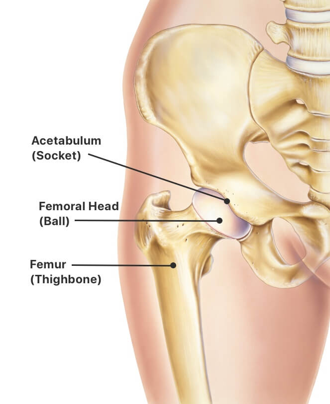
Hip Replacement Procedure Symptoms Types Of Implants And Risks from www.drugwatch.com When you stand or walk, all the weight of your upper body rests on them. The bones of the leg are the femur, tibia, fibula and patella. The foot bones shown in this diagram are the talus, navicular, cuneiform, cuboid, metatarsals and calcaneus. The hip itself is a ball and socket joint, much like the shoulder. The ilium, ischium, and the pubis. The knee joint is the largest joint in the body and is primarily a hinge joint, although some sliding and rotation occur. The ball and socket bony structure. By natalia kremenon january 21, 2021in wiring diagram231 views.
Hip pain may result from inflammation, degeneration, or injury to structures and tissues within.
Ankle and foot pain massage therapy connections. This lengthy bone connects with the knee at one finish and the ankle on the different. The piriformis muscle is what lets the hip rotate laterally, which is necessary in order for the legs to cross. There are numerous structures that contribute stability to the hip: The femur is the upper leg bone or thigh. The ilium bone forms the superior portion of the os coxa, the ischium bone the lower posterior portion, and the pubic bone (pubis) the lower anterior portion. The bones of the leg are the femur, tibia, fibula and patella. The hip joint is made up of two bones: The ilium, ischium, and the pubis. Bringing the leg back towards the midline. Start studying leg bone diagram. The ball and socket bony structure. Quizzes on human skeletal system anatomy, bone the musculoskeletal segment of the leg, including the foot bones (ankle, heel bone, toe bones), fibula and tibia, knee, femur and femoral neck, hip and sacrum.
Of the corollary to this is when pathology arising from the hip joint and structures around it manifests as upper leg bones diagram the junction of where these structures converge at the pubic bone. The bones of the leg are the femur, tibia, fibula and patella. Bones of right thigh and leg. Click and start learning now! The piriformis muscle is what lets the hip rotate laterally, which is necessary in order for the legs to cross.
Femur And Pelvic Bones Diagram Fusebox And Wiring Diagram Series Ton Series Ton Sirtarghe It from fpnotebook.com The bones of the leg are the femur, tibia, fibula and patella. Click and start learning now! These muscles include the adductors (adductor magnus. The bones together make up the hip. Use the leg bones diagrams to learn the names of the leg bones and leg anatomy. Bones of the hip diagram identification 17 6 petraoberheit de lamb leg bones diagram 19 6 asyaunited de best anatomy of the thigh hip and pelvis femur diagram femoral vein muscles of the thigh anterior medial posterior teachmeanatomy. On top of that layer of muscle is the iliotibial band, which starts at the brim of your pelvis outside the hip joint and runs down your leg. Hip and thigh bones joints muscles kenhub.
Femur bone diagram, picture of femur bone diagram.
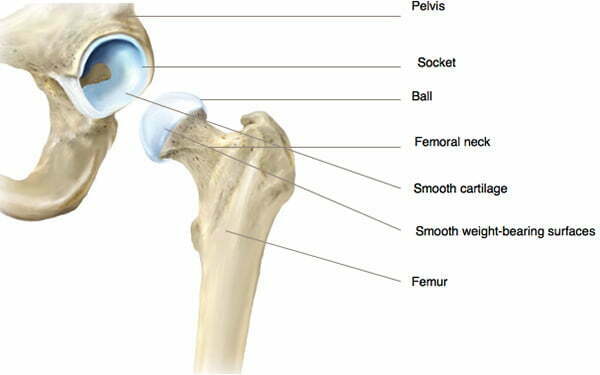
The pelvic bone area is different in men and women. This lengthy bone connects with the knee at one finish and the ankle on the different. The femur is the upper leg bone or thigh. Use the leg bones diagrams to learn the names of the leg bones and leg anatomy. Hip muscle strains info florida orthopaedic institute. Cited after worker's leg amputated. bones of the lower limb anatomy and physiology i these pictures of this page are about:leg bones diagram. This diagram depicts diagram leg bones anatomy. When you stand or walk, all the weight of your upper body rests on them. Want to learn more about it? Your leg bones are the longest and strongest bones in your body. Muscles of hip, thigh, leg, and foot. The piriformis muscle is what lets the hip rotate laterally, which is necessary in order for the legs to cross. The bone surfaces of the femoral head and acetabulum have a smooth durable layer of articular cartilage that cushions the ends of the bones and allows for smooth movement.
The hip itself is a ball and socket joint, much like the shoulder leg bone diagram. The bones of the leg are the femur, tibia, fibula and patella.


Posting Komentar
0 Komentar